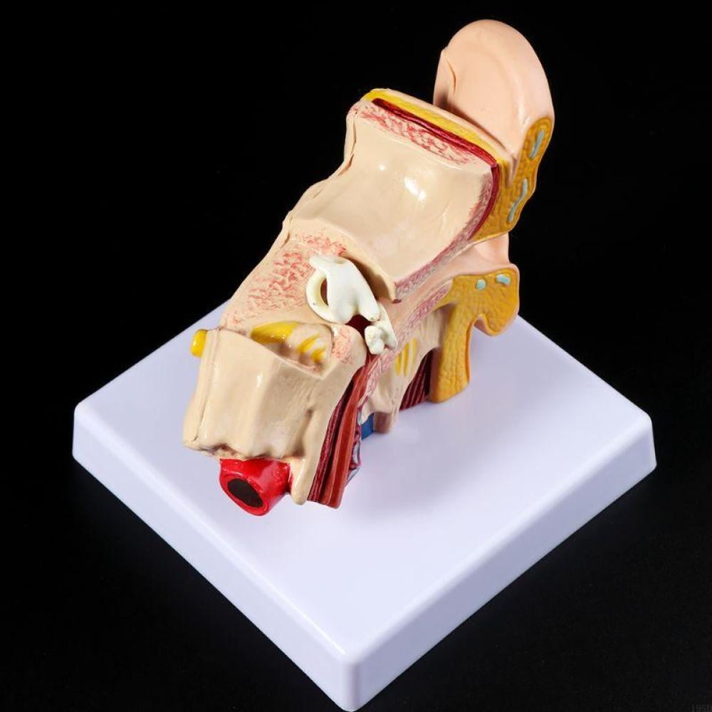
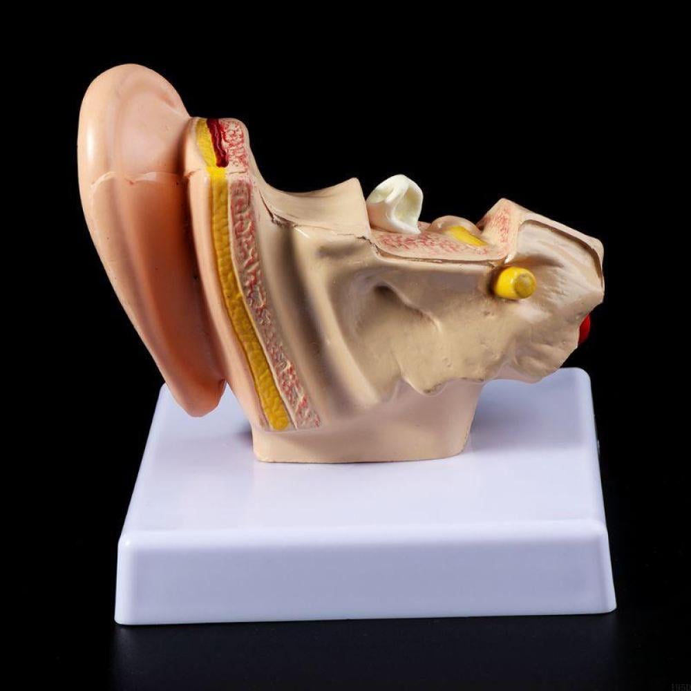
Magnified ear anatomy model (1.5x Life Size) – A deeper view of hearing and equilibrium details
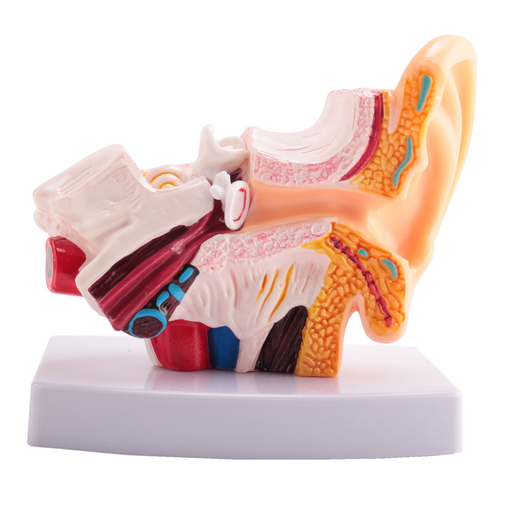
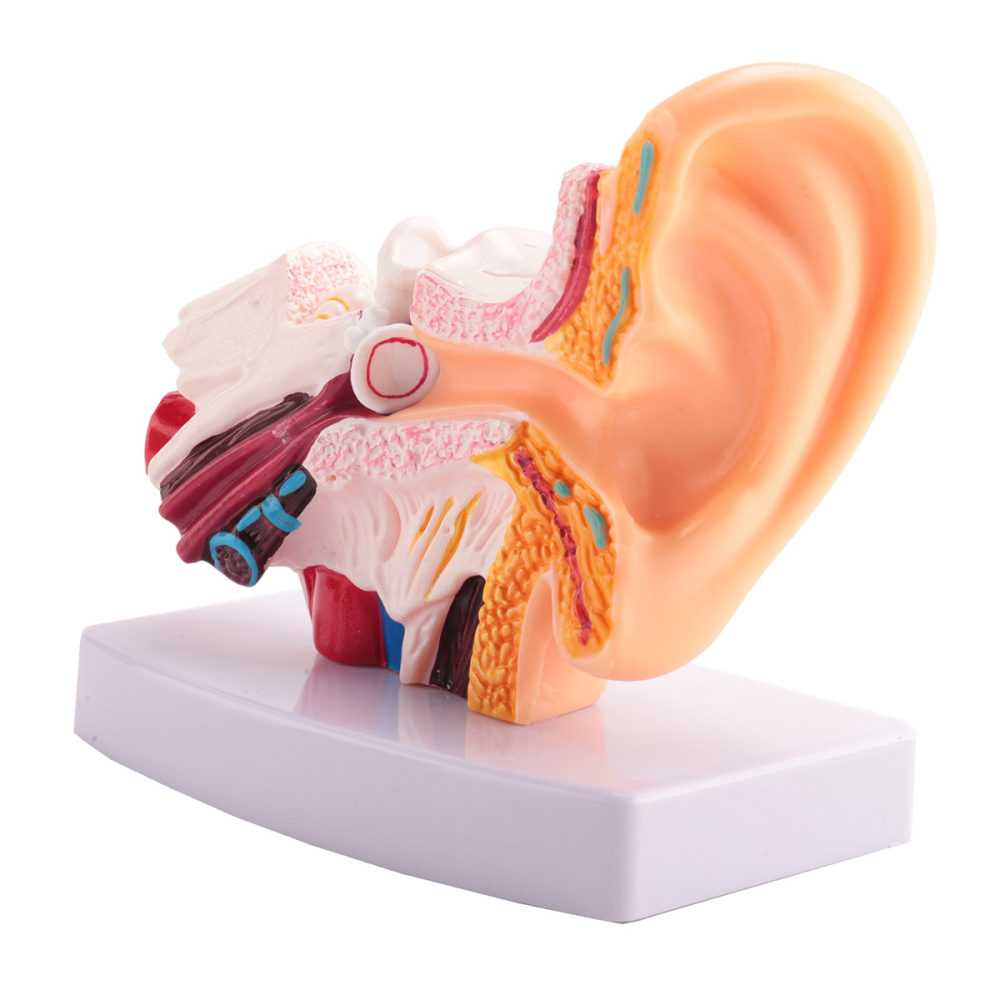
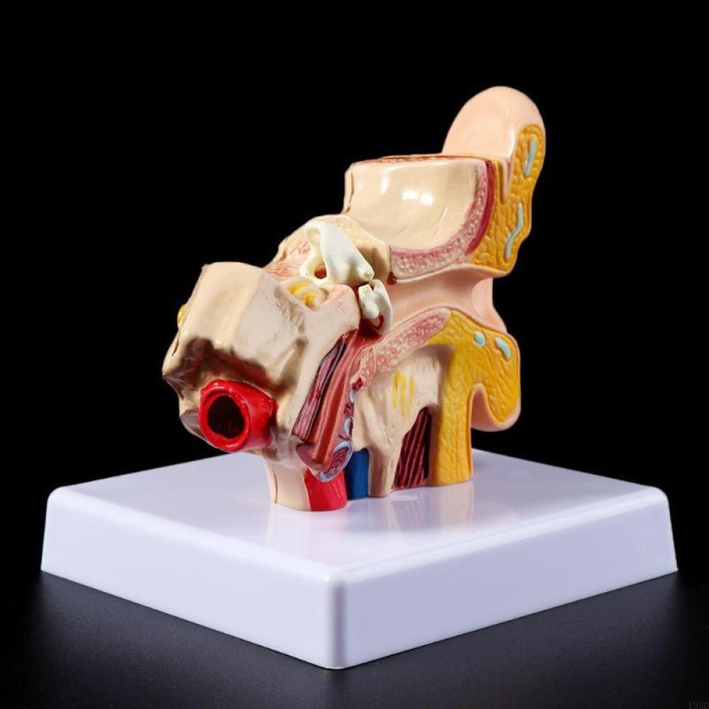
Because the human ear contains the smallest and most delicate bones and structures in the body, studying it at life-size can be challenging. Therefore, at Good Doctor, we offer a professional ear model magnified 1.5 times larger than life-size, giving you exceptional clarity in viewing the complex internal structures that are difficult to observe in standard models.
Why is this model a smart choice for your clinic or office?
- 3-part educational design: The model is designed to be disassembled into 3 main pieces, allowing you to explore the outer ear, middle ear (including eardrum and ossicles), and inner ear (cochlea and semicircular canals) separately and accurately.
- Superb clarity of detail: Thanks to 1.5x magnification, the model clearly highlights the three ossicles (malleus, incus, stapes), the auditory canal, and the auditory nerve, making it easier for students to memorize and for patients to explain.
- Durability and high quality (PVC): Made of strong and impact-resistant PVC material, designed to withstand frequent use in busy educational and medical office environments.
- An ideal communication tool: It helps ear, nose, and throat (ENT) doctors simplify complex concepts for patients, such as explaining ear perforations, middle ear infections, or balance problems.
What does the model illustrate?
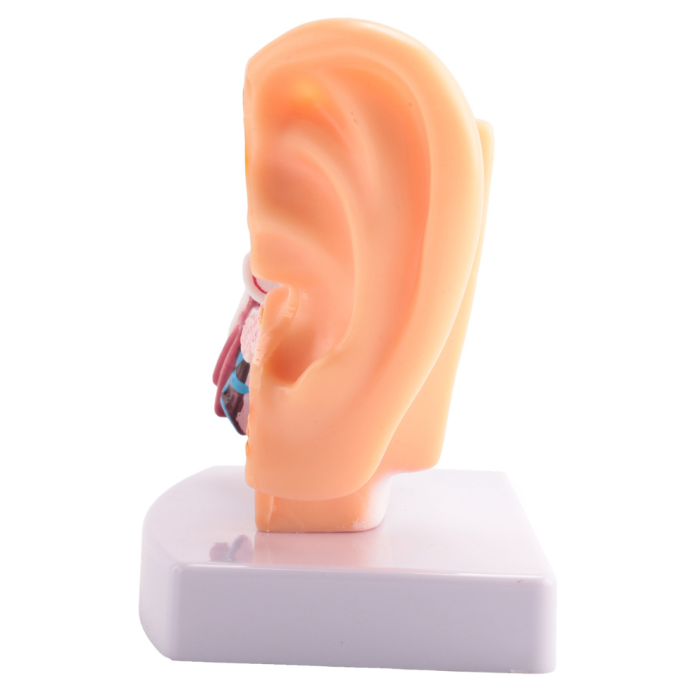
- The outer ear: the auricle and the external auditory canal.
- Middle ear: the eardrum, and the three auditory ossicles.
- Inner ear: the cochlea, semicircular canals, and vestibulocochlear nerve.
❓ 4. Frequently Asked Questions:
Q: What is the benefit of the model being magnified by 1.5 times? A: This carefully considered magnification makes the very small parts of the inner and middle ear visible to the naked eye without the need for magnifying lenses, making it the best tool for explaining clinical skills and anatomy.
Q: Can the eardrum and ossicles be removed? A: Yes, the model is designed to be interactive; you can remove certain parts to see the structures behind them, which enhances a deep understanding of the anatomical depth of the ear.
Q: Does the model come with a base? A: Yes, the model comes mounted on a sturdy base that allows it to be displayed stably and professionally on a desk or in a laboratory.
🔗 5. Related Products:
- Professional otoscope : The clinical instrument used to examine the live ear and compare it to a stereoscopic model.
- The complete OSCE Kit : To equip yourself with all the tools for clinical examination.
- 128 Hz Medical Tuning Fork : For clinical hearing tests related to ear anatomy.